
Methods of Preservation
Focusing on an array of preservative methods such as plastination, corrosion casting, wet specimen care, and cosmetic restorations.
Conservation of history
Since the earliest days of medicine, cadavers have been used in understanding anatomy and physiology. Originally each student of medicine was encouraged to own their own skeleton if they were expected to succeed. As history progressed the medical facilities themselves housed the specimens and supplied students with equal learning opportunities.
As preservation practices continued to progress the elongation of a specimen allowed for years to decades of use per each donated individual. The reason for decommissioning of learning cadavers are attributed to the amount of handle and invasive study on any given organ or remain. Once a specimen is too damaged for accurate teaching they were often taken home by doctors of medicine and housed in personal collections.
Nearly all medical remains that reside in private collections are sourced from doctors estates after the individuals passing - as well as retired facilities once used in processing and creating teaching models as we see them today.
Wet Specimen Conservation
-

Rehydration
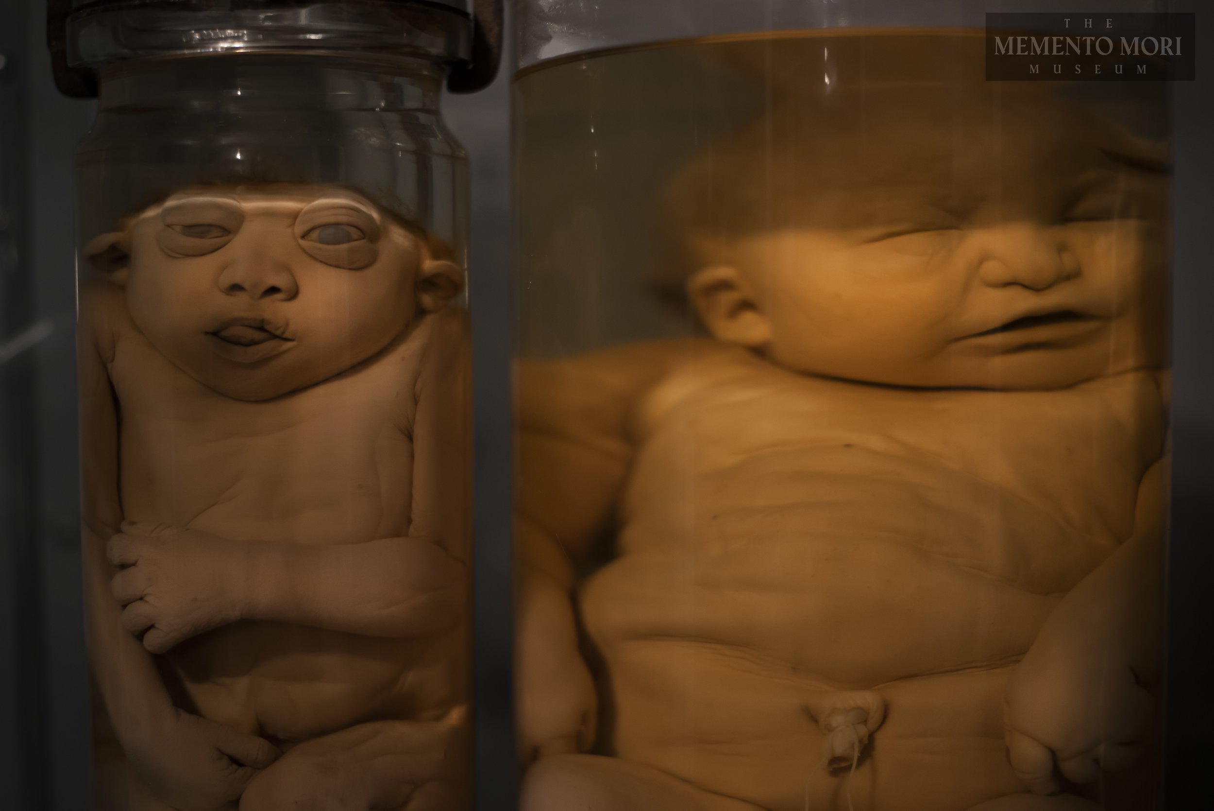
Many of the specimens seen today were originally preserved decades past. No matter the container used for housing, a significant amount of evaporation occurs, leading to dehydration and on occasion mummification. Understanding the chemical compositions of various preservatory procedures is important in the steps of purging chemical contents from the tissue, rehydrating, and stabilizing for restorative efforts.
-

Fluid Changes
The older the specimen, the more time natural purging of biological material and preservatory chemical reactions cloud the suspension agent. Oftentimes the liquids become a yellow, orange, or dark brown in color and presents with sediment formation on the bottom of the housing container. It is important that all medical specimens be cared for and liquids be replenished as evaporation occurs, to prevent any tissue having exposure to air, and to prevent degradation from sunlight and fluid expiration.
-

Stabilization
After the process of fluid removal and gentle purging of previous chemical contents from the tissue of the specimen, slow introduction to new preservatory agents follow. These processes can take many weeks so as not to shock biological material and cause further damage. Once established the specimen is stabilized in the full percentage of preservation liquids chosen, and proper airtight sealing of the container may commence, and the specimen is set until it’s next required fluid change.
Why do we need restoration and conservation?
Specimens that are graciously donated to science deserve the upmost respect and care - long after their immediate use in the scientific field. When specimens are deemed unusable they are oftentimes cremated or thrown away, in fact the majority of cadavers donated to science are not used in the medical field, but are sold for monetary gain by massive companies meant to collect donated bodies themselves. There are many articles available online on specific companies and their misuse of remains leading to the supply and demand for actual educational facilities abysmal.
As quoted in an article written by Reuters: “Medical school officials in Pennsylvania and Florida report that competition from Science Care and other brokers has reduced the number of bodies donated to schools to train students. Science Care markets itself more aggressively than medical schools, they say, and offers donors more favorable terms, such as picking up the body for free.”
Because of the twisted nature of these companies, Jeremy dedicates his time in preserving and reconditioning retired specimens to continue their use to students - sourcing from MD’s estate sales, medical facilities, and museum donations. Every body donated to science deserves to be used for it’s intended purpose, even if that requires months of conversative efforts to do so.

Continued efforts in education


Following Jeremys efforts in education and reconditioning of human remains, he is in continual collaboration with other dedicated artists and conservation specialists in the fight to reinstate donated remains in their intended use. A current collaborative project in restoration is the mounting and displays of retired medical bones to help prevent continued damage and encourage another “lifetime” of study.
All specimens that are mounted are done so by hand and the continual effort is made to create custom displays that maintain the integrity of the bones and provide documentations on their anatomy to help educate on their physiological use in the human body.
Specimens supplied by JLP and mounted by AnatomieTDN